Femur Fracture
What is a femur fracture?
Your thighbone is known as the femur bone. The femur spans from the hip to the knee. The femur is the longest and strongest bone in your body. Since femur bones are so strong, it usually takes a lot of force to break them. One of the most common causes of femur fractures (breaks) are car crashes.
A fracture is a break in the bone in 2 or more pieces. There are different types of femur fractures.
-
Femur fractures can be nondisplaced (bone is broken but the parts still line up correctly) or displaced (out of alignment).
-
Femur fractures can be closed (skin intact) or open (the bone has gone through the skin). Open femur fractures (also known as compound fractures) can happen when either pieces of the bone are sticking through the skin or a wound penetrates down to the broken bone. These types of fractures cause more damage to the nearby tissues (muscles, tendons, and ligaments), have a higher risk of complications (such as infections), and take a longer time to heal.
What parts of the femur can break?
Distal femur fracture
-
Description: This type of femur fracture is just above the knee joint where the bone flares out like an upside-down funnel. The distal femur makes up part of the knee joint. These fractures can sometimes extend into the knee joint and can separate the surface of the bone into multiple parts. These types of distal femur fractures are known as intra-articular and can be more difficult to treat. Distal femur fractures happen most often in younger people and the elderly.
-
Treatment: These types of fractures may require surgery, depending on how bad they are.
Femoral shaft fracture
|

|
Proximal femur fracture
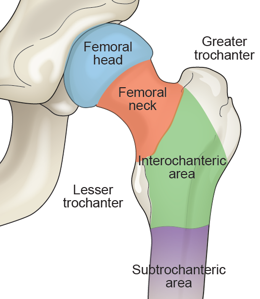
This type of femur fracture occurs close to the ball (femur head) of the femur that makes up part of the hip joint. These types of fractures happen most often in falls by older people with weakened bones. There are 4 types of proximal femur fractures. These are based on the location of the fracture.
1. Femoral neck fracture
2. Intertrochanteric area fracture
-
Description: This is the area below the neck of the femur and above the long part or the shaft. These fractures happen below the femoral neck. It is a bigger area between the greater and lesser trochanters.
-
Treatment: These fractures are often treated with a sliding compression hip screw and side plate or with an intramedullary nail (rod).
-
The compression hip screw is attached to the outer part of the bone with screws. Then a larger screw is placed through the plate and into the femoral head and neck.
-
The intramedullary nail is placed into the marrow canal (center) of the bone through an opening made at the greater trochanter. Then 1 or more screws are placed through the nail and into the femoral head.
3. Subtrochanteric area fracture
-
Description: This is the upper part of the shaft of the femur below the greater and lesser trochanters.
-
Treatment: These fractures are often treated with an intramedullary nail (rod). It is placed into the marrow canal (center) of the femur shaft. Then a screw is placed through the nail into the femoral head. Multiple screws may be placed at the lower end of the nail at the knee.
4. Femoral head fracture
-
Description: This is the ball of the femur that sits in the socket. These fractures are very rare.
-
Treatment: These fractures may be able to be treated without surgery. When treated with surgery, it is often by open reduction and internal fixation (ORIF) with screws.
Which image studies help diagnose femur fractures?
X-rays. This study provides images of dense structures like bones. X-rays are often the first scans done to find bone fractures.
Magnetic Resonance Imaging (MRI) scans. This study provides images of soft tissue structures and bone. MRIs are very sensitive and can sometimes detect a small or incomplete fracture that cannot be seen on an X-ray.
Computerized Tomography (CT) scans. This study provides a detailed cross-sectional image of the bone. CT scans are often ordered for surgeons to get more detailed imaging of a specific fracture.